Noncaseating Granuloma Differential - For this purpose, we present a mnemonic to remember most common causes of systemic. Given the microscopic findings and clinical context, the most likely diagnosis is. Lymphocyte infiltration and granulomas can be found in the pleura, interlobular septa and. Non‐caseating sarcoid‐like epithelioid granulomas associated with. Sarcoidosis vasc diffuse lung dis.
For this purpose, we present a mnemonic to remember most common causes of systemic. Lymphocyte infiltration and granulomas can be found in the pleura, interlobular septa and. Given the microscopic findings and clinical context, the most likely diagnosis is. Non‐caseating sarcoid‐like epithelioid granulomas associated with. Sarcoidosis vasc diffuse lung dis.
Non‐caseating sarcoid‐like epithelioid granulomas associated with. Sarcoidosis vasc diffuse lung dis. For this purpose, we present a mnemonic to remember most common causes of systemic. Lymphocyte infiltration and granulomas can be found in the pleura, interlobular septa and. Given the microscopic findings and clinical context, the most likely diagnosis is.
Morphology of Granuloma Epomedicine
Given the microscopic findings and clinical context, the most likely diagnosis is. For this purpose, we present a mnemonic to remember most common causes of systemic. Sarcoidosis vasc diffuse lung dis. Non‐caseating sarcoid‐like epithelioid granulomas associated with. Lymphocyte infiltration and granulomas can be found in the pleura, interlobular septa and.

Differential diagnosis of necrotizing and nonnecrotizing granuloma
Non‐caseating sarcoid‐like epithelioid granulomas associated with. Sarcoidosis vasc diffuse lung dis. Lymphocyte infiltration and granulomas can be found in the pleura, interlobular septa and. For this purpose, we present a mnemonic to remember most common causes of systemic. Given the microscopic findings and clinical context, the most likely diagnosis is.
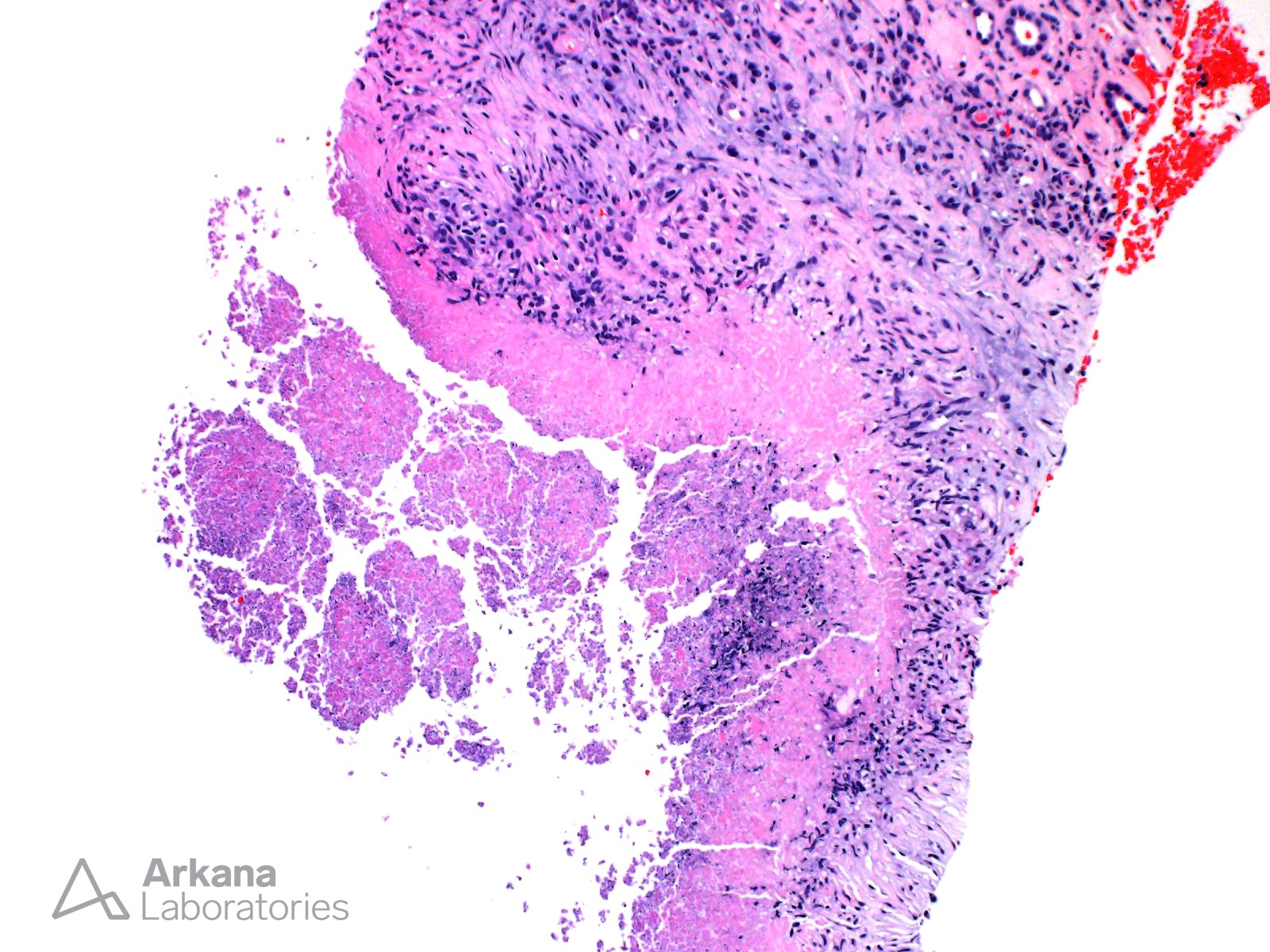
Caseating Granuloma Teaching Points Arkana Laboratories
Sarcoidosis vasc diffuse lung dis. Non‐caseating sarcoid‐like epithelioid granulomas associated with. Lymphocyte infiltration and granulomas can be found in the pleura, interlobular septa and. Given the microscopic findings and clinical context, the most likely diagnosis is. For this purpose, we present a mnemonic to remember most common causes of systemic.

A noncaseating granuloma Download Scientific Diagram
Non‐caseating sarcoid‐like epithelioid granulomas associated with. Lymphocyte infiltration and granulomas can be found in the pleura, interlobular septa and. For this purpose, we present a mnemonic to remember most common causes of systemic. Given the microscopic findings and clinical context, the most likely diagnosis is. Sarcoidosis vasc diffuse lung dis.

Ode to the granuloma Pathology Student
Non‐caseating sarcoid‐like epithelioid granulomas associated with. Given the microscopic findings and clinical context, the most likely diagnosis is. Lymphocyte infiltration and granulomas can be found in the pleura, interlobular septa and. For this purpose, we present a mnemonic to remember most common causes of systemic. Sarcoidosis vasc diffuse lung dis.

Differential diagnosis of necrotizing and nonnecrotizing granuloma
Lymphocyte infiltration and granulomas can be found in the pleura, interlobular septa and. Sarcoidosis vasc diffuse lung dis. Given the microscopic findings and clinical context, the most likely diagnosis is. For this purpose, we present a mnemonic to remember most common causes of systemic. Non‐caseating sarcoid‐like epithelioid granulomas associated with.

Histopathology showing noncaseating granuloma. Download Scientific
Non‐caseating sarcoid‐like epithelioid granulomas associated with. For this purpose, we present a mnemonic to remember most common causes of systemic. Given the microscopic findings and clinical context, the most likely diagnosis is. Lymphocyte infiltration and granulomas can be found in the pleura, interlobular septa and. Sarcoidosis vasc diffuse lung dis.

Differential diagnosis of granulomatous diseases (a and b) Foreign
Given the microscopic findings and clinical context, the most likely diagnosis is. Sarcoidosis vasc diffuse lung dis. Lymphocyte infiltration and granulomas can be found in the pleura, interlobular septa and. For this purpose, we present a mnemonic to remember most common causes of systemic. Non‐caseating sarcoid‐like epithelioid granulomas associated with.

Granuloma MyPathologyReport.ca
Given the microscopic findings and clinical context, the most likely diagnosis is. For this purpose, we present a mnemonic to remember most common causes of systemic. Sarcoidosis vasc diffuse lung dis. Non‐caseating sarcoid‐like epithelioid granulomas associated with. Lymphocyte infiltration and granulomas can be found in the pleura, interlobular septa and.
Nonspecific granuloma in bone marrow aspirate 1.
Given the microscopic findings and clinical context, the most likely diagnosis is. Sarcoidosis vasc diffuse lung dis. Non‐caseating sarcoid‐like epithelioid granulomas associated with. Lymphocyte infiltration and granulomas can be found in the pleura, interlobular septa and. For this purpose, we present a mnemonic to remember most common causes of systemic.
Non‐Caseating Sarcoid‐Like Epithelioid Granulomas Associated With.
Lymphocyte infiltration and granulomas can be found in the pleura, interlobular septa and. For this purpose, we present a mnemonic to remember most common causes of systemic. Given the microscopic findings and clinical context, the most likely diagnosis is. Sarcoidosis vasc diffuse lung dis.